1. K-State home
  2. »NICKS
  3. »Electron Microscopy Facility

Nanotechnology Innovation Center of Kansas State

Electron Microscopy Facility

The Nanotechnology Innovation Center of Kansas State (NICKS) hosts an electron microscopy (EM) facility located within the College of Veterinary Medicine on Denison Avenue. This facility can be used to help improve the scientific output of Kansas State University researchers, while providing faculty, students and staff with guidance for EM research.

The facility can conduct both transmission electron microscopy (TEM) and a scanning electron microscopy (SEM).

  • For TEM, there is a FEI Tecnai G2 Spirit BioTWIN that provides a 0.34 nm resolution, 120Kv and a GATAN digital imaging system with a HAADF detector for STEM imaging and an Oxford detector <135 eV for elemental analysis.
  • For SEM, there is a Hitachi S-3500N scope that provides a 3.0 nm resolution and also equipped with an Oxford detector <135 eV for elemental analysis.

“Our new EM facility has all the necessary equipment for proper sample fixation, processing, sectioning and visualization for TEM and SEM,” said Dr. Nancy Monteiro-Riviere, university distinguished professor and director of NICKS. “The EM facility also offers analysis for a variety of samples from biological tissues, body fluid, bacteria, viruses and samples from other scientific fields. Applications include cell biology, structural biology, soft matter and nanomaterials, nanoparticles and other fields of nanotechnology where one requires nano level imaging.”

Specific technical expertise is listed below:

  • Study of morphological changes in cells and organelles under certain disease or experimental conditions. Viral and/or bacterial deposits under infectious conditions in animals and/or plant tissue, bacterial, fungal, algae, cultured cell lines, and artificial cell matrices.
  • Subcellular localization of pathology related protein or interested biomolecules inside the cell using immunolabeling of ultrathin frozen and plastic embedded sections.
  • Elemental analysis of metal accumulation inside cells using X-ray dispersive spectroscopy.
  • Provides analytical support for characterization (DLS, Zeta measurements) and interaction of nanoparticles in cells, tissues and biological fluids for drug delivery. The NICKS Center has other specific centralized instrumentation needed for nano research which is also available.
  • Protein dynamics such as protein miss-folding, aggregation, amyloid formation, protein corona complex and protein interactions with nanoparticles and other molecules, Liposomes and lipid self-assembled nanostructures and other biological material using negative staining.
  • Nanocharacterization, an elemental analysis using STEM and X-ray dispersive spectroscopy of metallic nanoparticles, polymeric nanoparticles, core shell nanoparticles and soft polymer nanocomposites like soft polymer laced with nanostructures like carbon nanotubes, fullerenes, quantum dot nanoparticles and other nanostructured materials.

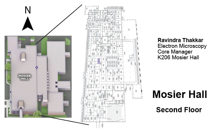
Dr. Monteiro-Riviere noted that the EM microscopy facility has the capability for cryo electron microscopy studies.

The EM microscopy facility is scheduled to have the capability for cryo electron microscopy studies. “The TEM is cryo-compatible and equipped with a low-dose mode for beam-sensitive vitrified sample observation at liquid nitrogen temperatures,” she said. “We still need a few more additional attachments before we can offer full cryo capabilities. If faculty are interested in using cryo for their research, please let us know so we can outfit the scope with the proper cryo attachments.”

NICKS offers consultation services concerning technical methods, budgeting, SOP preparation and equipment selection.  Please contact the EM manager Ravi Thakkarravithakkar@vet.k-state.edu